
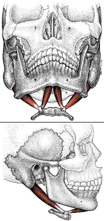
The digastric is a double muscle of the throat which is located under the chin, behind and below the corner of the jaw, immediately in front of the top of the sternocleidomastoid, one for each side of the jaw and neck. It gets its name from the Greek word for “two bellies”. The Greek word dia means double and gaster means belly hence digastric meaning “two-bellied”. The digastric is made up of an anterior and posterior belly. The anterior belly extends from the digastric fossa of the mandible and the posterior belly extends from the mastoid notch of the temporal bone. Both bellies then insert to the body of the hyoid bone via a fibrous loop over a common intermediate tendon between the two bellies.
The digastric assists the lateral pterygoid in depressing the mandible (opening the jaw), primarily during maximum depression or very quick forceful opening of the mouth. Together with the stylohyoid, geniohyoid, and mylohyoid muscles, it is known as a suprahyoid muscle and these other hyoid muscles are also its synergist in assisting in mandible depression, though the lateral pterygoid is the more important muscle in this action. The suprahyoid muscles, in general, elevate the hyoid bone and are important for control of the esophagus and pharynx during swallowing and speaking.
Overwork occurs in the digastric usually due to the pressure of an overactive masseter accompanied by habitual open mouth breathing due to sinus problems or secondary to bruxism.
Digastric Origin, Insertion and Action and Synergists
Anterior Belly
Origin: Digastric Fossa of Mandible, close to symphysis of mandible.
Insertion: Both bellies united by a common intermediate tendon, attaching to hyoid bone through a fibrous loop.
Action: Elevates hyoid bone, depresses mandible to open mouth, assists retruding mandible.
Synergists: For mandible depression: Lateral pterygoid (for maximum jaw opening) and infrayoid muscles; For mandible retrusion: Posterior temporalis fibers and masseter.
Antagonists: To jaw opening: the mandible elevators including the masseter, temporalis, medial pterygoid and superior lateral pterygoid.
Posterior Belly
Origin: Mastoid Notch of Temporal Bone under attachments of longissimus capitis, splenius capitus and sternocleidomastoid.
Insertion: Same as anterior belly.
Action: Same as anterior belly.
Synergists: Same as anterior.
Antagonists: Same as anterior. 1Arnold, M. A. “Arnold’s Glossary of Anatomy.” Discipline of Anatomy and Histology – The University of Sydney. Web. 21 Nov. 2010. <http://www.anatomy.usyd.edu.au/glossary/>.,2Simons, David G., Janet G. Travell, Lois S. Simons, and Janet G. Travell. “Chp. 12: Digastric Muscle.” Travell & Simons’ Myofascial Pain and Dysfunction: the Trigger Point Manual. Baltimore: Williams & Wilkins, 1999. 273-280. Print.
Sources
| ↲1 | Arnold, M. A. “Arnold’s Glossary of Anatomy.” Discipline of Anatomy and Histology – The University of Sydney. Web. 21 Nov. 2010. <http://www.anatomy.usyd.edu.au/glossary/>. |
|---|---|
| ↲2 | Simons, David G., Janet G. Travell, Lois S. Simons, and Janet G. Travell. “Chp. 12: Digastric Muscle.” Travell & Simons’ Myofascial Pain and Dysfunction: the Trigger Point Manual. Baltimore: Williams & Wilkins, 1999. 273-280. Print. |